International Week For Breast Feeding

courtsey image WBW
courtsey image WBW

Breast feeding

The world breast feeding week (WBW) was in the first week of august.

Yes my blog did not commemorate the event, simply because I realize that ground reality varies.

Most women today are aware of the need to nurse their child. But challenges are many. The woman could be working in the formal, or non-formal or home setting but she should be empowered to claim her and her baby’s right to be breast fed.

In 1993 WBW had a campaign on Mother-Friendly workplace initiative.  There has been viable improvement achieved over the past 22 yrs. There are laws in place and there are women who are not satisfied with the law. There are also women who abuse the law.

The 1990 Innocenti Declaration recognized that breast feeding provides ideal nutrition for the infants and contributes to their health, growth and development. There is much that remains to be done despite 25yrs of hard work and the progress achieved.
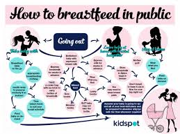
image courtesy WBW
image courtesy WBW

What WABA called for this year was

  • Organized global action to support such that they combine breast feeding and work is it in the formal sector or non-formal sector at home.
  • Maternity protection laws along the ILO Maternity protection convention.
  • Inclusion of breastfeeding target indicators in the sustainable development goals.

What I would like to share is what some women did achieve and this is what was shared on the platform of SHEROES.IN, when women came up with their job challenges.

Let me start with Dr.Lathika (name changed to maintain her privacy) she was heading the department of Periodontology  and was a nursing mother, first week after she returned to work post partum she would drive home nurse the kid and return. After which she just shifted the crib to her cabin. The child would there and she would excuse herself when she had to nurse him. When a staff nurse was in the similar situation she suggested that the nurse use either uses the nurses room, she then ensured that there was a duty staff room, like the students lounge where duty doctors could relax and many of the lady staff started leaving their kids in that room along with the baby sitter.

Mrs.Shashi Deshpande (name changed to ensure her privacy) taught in an engineering college. Post maternity leave, she would express milk into a feeding bottle and refrigerate it, and her mother would feed the child while she was away. Of course when she was at home she nursed the child herself.
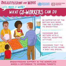
Image courtesy internet

But the most interesting share came from the CEO of facebook, when she had to travel  leaving a month old child was definitely  not done, definitely for two months period. Colleagues suggested using fed-ex to transport expressed milk, various other suggestion each seemed more unviable than the other. She then came up with the simplest solution. She requested for a place within her workspace where she could leave the child with a sitter and take a break to feed. To her surprise the other person on her team was a single father with three months old kid, things worked fine.

At the end the end of the day generations of women have coped with work and nursing. We just need to tap that memory and rewrite it in contemporary context.

“Breastfeeding does not have to be hard. Breastfeeding is natural. With rare exceptions, it becomes hard only because of all the interference caused by the medicalization of birth and unsupportive culture. Animal’s breastfeed instinctively with no need for supplementation, classes, or support. We as humans also have these instincts. We have become so disconnected. Breastfeeding my children has been one of my greatest joys in life, and I am filled with sorrow when I imagine how many mothers and infants haven’t been able to experience this because of misinformation.”
― Adrienne CarmackReclaiming My Birth Rights

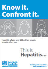
Hepatitis awarenes


hepatitisWorld Hepatitis day July 28th.

Hepatitis is the inflammation of the liver. The condition is self-limiting but sometimes process to scarring called cirrhosis of the liver.  It can also progress to liver cancer.

The most common cause of hepatitis in the world is virus. Through infection, toxicities like alcohol, drugs and some autoimmune diseases can also cause this condition.

Hepatitis could be type A,B, C,D and E. These 5 types are of greatest concern, because they are potential epidemic.

Hepatitis A and E are typically caused by ingestion of contaminated food or water, while B, C and D usually occur as a result of parental contact with infected body fluids. This could happen due to contaminated medical equipments, mother to child at birth, sexual contact or contaminated equipment.

Symptoms of acute infection occurs as jaundice (yellowing of skin and eyes), dark urine, extreme fatigue nausea, vomiting and abdominal pain.

Different hepatitis identified by A, B, C, and E. All these cause liver disease, though vary in important ways.

Hepatitis A Virus (HAV) is present in the faeces of an infected person. This gets transmitted through contaminated water or food. It can spread through certain sexual practises too. If the infection is mild the person recovers and acquires immunity to the strain. It can be severe and life threatening though. The outbreak of this strain is seen in places where sanitation is poor.—vaccines are available for this.

Hepatitis B Virus (HBV) is transmitted through exposure to infected blood, semen and other body fluids. HBV can be transmitted from infected mothers to infants at the time of birth or from family member to infant in early childhood. Transmission may also occur through transfusions of HBV- contaminated blood and blood products or medical procedural equipment. Health care workers are also at the risk of HBV.

Hepatitis C Virus (HCV) is mostly transmitted through exposure of infected blood. This is transmitted through blood, body fluids, and contaminated medical equipments. Sexual transmission is also possible but it’s less common. There is vaccine for HCV.

Hepatitis D Virus (HDV) infections occur only in those who are infected with HBV. The dual infection of HDV and HBV can result in more serious disease and worse outcome. Hepatitis B vaccines provide protection from HDV infection.

Hepatitis e virus (HEV) is transmitted through consumption of contaminated water or food. HEV is a common cause of hepatitis outbreaks in developing parts of the world and is increasingly recognized an important cause of disease developed countries. Safe and effective vaccines to prevent HEV infection have been developed though not widely available.

Hepatitis awareness–Hepatitis C

hepatitisWorld Hepatitis awareness Hepatitis C

July 28th is the world Hepatitis awareness day.

The Hepatitis C virus causes both acute and chronic infection.

Acute is something that is happens quickly, it is usually asymptomatic and rarely life-threatening. About 15-45% of the infected people spontaneous heal within 6 months and do not require treatment. The other 55-85%  go into the chronic stage. Of whom 15-30% could develop into cirrhosis of the liver in 20 yrs.

Geographically hepatitis C is found world wide. Though central and east Asia and northern Africa are most affected. The epidemic  could be concentrated in high risk populations like people use infected needles, or could be general. There are multiple strains of HCV virus and their distribution.

Transmission  is blood borne. And common pathways are

  • Use of infected needles.
  • Use of contaminated medical equipment without adequate sterilization.
  • Transmission of contaminated blood.
  • Transmission through body fluids and from mother to her fetus these are however rare.

Hepatitis however does not spread through breast milk, food, water or casual contact like hugging or kissing or share food with an infected person.

Symptoms  show after a period of 2 weeks or 6 months of incubation. About 80% of the people do not exhibit any symptoms. Those do exhibit acute symptoms may show fever, fatigue, decreased appetite  nausea, vomiting, abdominal pain, dark urine, grey-colored faeces, joint pain and jaundice.(jaundice is the yellowing of skin and whites of the eyes.)

Screening and diagnosis of the early stage is rare, as it is asymptomatic.  Many a times it remains undiagnosed until serious liver damage has developed.

The diagnosis of HCV is in 2 steps.

  • Screening for anti HCV antibodies with a serological test – this identifies people who have been infected with virus.
  • If the serological test is positive, a nucleic aci test for HCV-RNA  is needed to confirm  chronic strong immune response without the need for treatment. Although no longer infected they will still test positive for anti-HCV  infection  because 15-45% of the people infected with HCV spontaneously clear the infection.

Once the person is diagnosed of harboring hepatitis C the liver is damaged is assessed for fibrosis and cirrhosis. This can be done by non-invasive test. The treatment plan and disease management is decided after identifying the strain. There are 6 strains of HCV.

Getting tested:

Early diagnosis can prevent further destruction  from the disease and transmission of the virus. high risk population could periodically be screened for the infection. And high risk include

  • People who inject drugs.
  • Recipients of blood products from probable unsafe source.
  • People undergo going invasive health care procedures in healthcare facilities with inadequate infection control practices.
  • People who sexual partners of HCV infected people.
  • People using intranasal drugs.
  • People who have had tattoos or piercing.

Hepatitis C does not always require treatment as the immune system in most people can clear the infection. When necessary the goal of the treatment is to cure. The cure rate dependents on factors like the strain of virus and treatment given. The appropriate approach of treatment is decided after carefully  screening the patient.  The current standard treatment is an antiviral therapy which is a combination f interferon and ribavirin.  This combination is effective against all the strains.  Unfortunately this is poorly tolerated by in some patients and is not widely available globally either. this makes management of the treatment a little complex, many patients do not finish their treatment.

Scientific advances have lead to the development of new antiviral drugs for hepatitis C which is much more effective, safer and better tolerated than existing therapies. These therapies are known as oral directly acting antiviral agents. Therapies simplify hepatitis C by significantly decreasing monitoring requirements and by increasing cure rates.  Though the production cost of DAA is low, the intial price set by the pharmaceutical companies are high and access to these drugs could be difficult even in high income countries.

WHO is launching new guidelines for screening care and treatment of persons with hepatitis C in April 2014.  These are the first guidelines dealing with hepatitis C treatment produced by WHO and complement the existing guidance on prevention of transmission of blood borne diseases in including HCV.

These guidelines are intended for the policy makers, government officials and others working in low-and middle income countries who are developing programs for the screening, care and treatment of persons with HCV infections. These guidelines will help expand of  treatment services to patients with HCV infections as they provide key recommendations in these areas and discuss considerations for implementations.

Prevention of HCV is on three planes, primary, secondary and tertiary. There are no vaccines for HCV so primary prevention of infection depends on  reduction of the risk of exposure to the virus, in healthcare settings, and high risk populations,  here are some examples of the primary prevention interventions recommended by WHO

  • Hand hygiene—including surgical hand preparation, hand washing and use of gloves.
  • Safe handling and disposal of sharps and waste.
  • Safe cleaning of equipment
  • Testing of donated blood
  • Improved access to safe blood.
  • Training of health personnel.

Secondary and tertiary prevention for infected with the HCV WHO recommends-

  • Education and counselling on options for care and treatment.
  • Immunization with the hepatitis A and B vaccines to prevent co-infection from hepatitis viruses to protect their liver.
  • Early and appropriate medical management and administration of antiviral therapy if appropriate.
  • Regular monitoring for early diagnosis of chronic liver disease.

WHO is working in the following areas to prevent and control viral hepatitis.

  • Raising awareness, promoting partnerships and mobilizing resources.
  • Formulating evidence based policy and data for action.
  • Prevention of transmission
  • Executing ,screening, care and treatment.

Hepatitis C is a liver disease caused by hepatitis C virus it can be both acute and chronic, and can last from a few weeks to a serious lifelong disease. It is blood borne. Significant number of those who suffer from chronic infection  develop liver cirrhosis or cancer. About 350,000-500,000 people die each from HCV.  This is curable 50-90%

though diagnosis and accessibility to the treatment is low. The success rate is  5o0-90% though research is on for vaccine against HCV.

Insulin Inhalation.

The inhalation of insulin into the lungs offers a new method of insulin treatment delivery for people with diabetes. The same features that make the lung so well suited for gas exchange also make it an ideal organ for absorption of small molecules into the bloodstream. The pulmonary alveolar surface area of the lung is 130 m2, the size of a tennis court, and the pulmonary capillary surface area is nearly as large at 115 m2. With each breath, air flows into nearly 300 million alveoli. Moreover, the alveolar lining cell is just 1 to 2 mcm from the pulmonary capillary lumen, a distance that favors rapid uptake into the bloodstream. Absorption of a molecule across the alveolar-capillary interface is inversely related to its molecular mass. Small peptides, such as insulin (approximately 6000 Daltons) are readily absorbed across the very thin, vesiculated, permeable membrane. Molecules that make it to the alveolar level have longer residence time there, because mucociliary mechanisms at this level are minimal. There are several factors affecting lower respiratory deposition of an aerosol or dry powder formulation. One of these is particle size. Particles greater than 5 mcm in diameter impact and are deposited in the pharynx and large airways. Particles 1 to 3 mcm generally reach the lower airways and alveoli. Particle velocity also affects deposition. Flow rates > 35 L/min or < 10 L/min will favor upper airway impaction, while flow rates of 15 to 25 L/min are ideal for lower airway deposition. Even under the best of circumstances, however, only the minority of an aerosol or dry powder usually makes it deep into the lungs Inhaled Human Insulin There are several forms of inhaled insulin, either approved or in development. The only inhaled insulin that is approved is Exubera® (insulin human [rDNA origin]) Inhalation Powder. Other forms in development include AERx (NovoNordisk), AIR (Lilly), Spiro (Dura), Technosphere Insulin (MannKind), and Aerodose (Aerogen). Some are not powder but aerosol. Their excipients and medication delivery systems also differ. Exubera is a dry powder insulin contained in small blisters of 1 mg and 3 mg potency. After the blister is inserted into the base of the inhaler, a vacuum is established by cocking the lever at the base, allowing aerosolization of the powder. The aerosolized particles are then inhaled.[21] Each actuation of the Exubera inhaler produces 200 mL of a homogeneous powder. This powder contains human (rDNA origin) insulin, and the excipients sodium citrate, glycine, sodium hydroxide (to maintain pH), and mannitol. None of these are known to be immunogenic. Mannitol has been clinically used as an agent in bronchoprovocation testing, but its concentration in Exubera is lower than the lowest dose used in such testing. There are several forms of inhaled insulin, either approved or in development. The only inhaled insulin that is approved is Exubera® (insulin human [rDNA origin]) Inhalation Powder. Other forms in development include AERx (NovoNordisk), AIR (Lilly), Spiro (Dura), Technosphere Insulin (MannKind), and Aerodose (Aerogen). Some are not powder but aerosol. Their excipients and medication delivery systems also differ. There are several forms of inhaled insulin, either approved or in development. The only inhaled insulin that is approved is Exubera® (insulin human [rDNA origin]) Inhalation Powder. Other forms in development include AERx (NovoNordisk), AIR (Lilly), Spiro (Dura), Technosphere Insulin (MannKind), and Aerodose (Aerogen). Some are not powder but aerosol. Their excipients and medication delivery systems also differ. Exubera is a dry powder insulin contained in small blisters of 1 mg and 3 mg potency. After the blister is inserted into the base of the inhaler, a vacuum is established by cocking the lever at the base, allowing aerosolization of the powder. The aerosolized particles are then inhaled.[21] Each actuation of the Exubera inhaler produces 200 mL of a homogeneous powder. This powder contains human (rDNA origin) insulin, and the excipients sodium citrate, glycine, sodium hydroxide (to maintain pH), and mannitol. None of these are known to be immunogenic. Mannitol has been clinically used as an agent in bronchoprovocation testing, but its concentration in Exubera is lower than the lowest dose used in such testing. The dose of Exubera that reaches the alveolar level is the “fine particle dose,” which consists of particles 3.3 mcm in diameter or smaller. The “fill mass” is the amount of insulin plus excipient in the individual insulin blister. In the case of a 1-mg Exubera blister, there is 0.7 mg of excipient and 1 mg of insulin; on actuation of the inhaler, 0.53 mg of insulin is emitted, of which 0.4 mg is less than 3.3 mcm in diameter. Thus, of the 1-mg insulin in the blister, 0.4 mg or 40% is deposited at the alveolar level. A 3-mg Exubera blister creates a 1.0 mg fine particle dose; hence, 33% is deposited in the alveoli. This explains why 3 1-mg blisters deliver more insulin (1.2 mg) than 1 3-mg blister, which delivers only 1 mg to the alveoli.[21] Most of the insulin that reaches the distal lung is absorbed. There is no evidence of insulin accumulation in the alveoli. The insulin that is not absorbed undergoes metabolic degradation or slow mucociliary clearance Smoking. Insulin absorption studies have looked at the maximum concentration of serum insulin after a dose of insulin (Cmax) as well as the area under the serum insulin vs. time curve (AUC) following that dose. Such studies have shown that active smoking increases absorption of inhaled insulin 2- to 5-fold. The mechanism by which smoking affects inhaled insulin absorption is still unknown. The absorption of subcutaneous insulin is not affected by smoking.[22] Cessation of smoking is accompanied by a reduction in absorption of inhaled insulin toward normal in as little as days. After a week of abstinence from smoking, inhaled insulin absorption decreases toward normal by as much as 50%. Resumption of smoking for just 3 days increases absorption of the peptide toward levels seen during chronic smoking.[22] Because of the wide variations in absorption of inhaled insulin observed with smoking, cigarette smoking within the past 6 months has been a contraindication to inhaled insulin use in Phase 2 and 3 studies, and remains a contraindication to use of inhaled insulin.[21] Passive smoking has been examined experimentally, in a study in which subjects were exposed to smoke in a smoking chamber for 2 hours at concentrations mimicking those found in a smoky bar. Contrary to active smoking, passive smoking appears to decrease inhaled insulin absorption by as much as 20% to 30%.[23] It is not clear how long this effect lasts after subjects are removed from the smoky environment. It is likewise unclear why passive smoking and active smoking have opposite effects on the absorption of inhaled insulin. Asthma, Chronic Obstructive Pulmonary Disease, and Their Treatment. Studies have shown that insulin absorption, as measured by area under the curve (AUC) and maximum concentration (Cmax), is 20% to 50% lower in mild to moderate asthmatics than in normals.[23] In contradistinction to asthmatics, a small cohort of patients with chronic bronchitis and emphysema having a predicted baseline forced expiratory volume in 1 second (FEV1) of 35% to 40% were tested and found to have rates of absorption for inhaled insulin that were 2-fold higher than those of subjects without chronic obstructive pulmonary disease (COPD).[23] Although both asthma and COPD are characterized by small airway inflammation, it is not known why inhaled insulin absorption is affected in opposite ways by these disorders. In patients with mild (FEV180% or more) or moderate (FEV180% or less) asthma, the administration of albuterol 30 minutes before taking inhaled insulin resulted in a 25% to 50% increase in systemic insulin absorption compared with administration of inhaled insulin alone. (Data on file) Inhaled fluticasone appears to have no effect on inhaled insulin absorption, but other medications used for asthma and COPD have not been systematically This slide summarizes how respiratory factors affect inhaled insulin absorption. Other Factors Affecting Absorption. In clinical studies of Exubera, episodes of viral upper respiratory infection, laryngitis, and acute bronchitis had no discernible effect in either direction on inhaled insulin absorption. There were not enough cases of pneumonia in phase 2/3 studies to be able to comment on the effects of a more severe infection on inhaled Respiratory Tract Effects of Inhaled Insulin Symptoms, Adverse Events. Respiratory safety of inhaled insulin has been a concern for several reasons: 1. It is a novel drug substance with novel excipients that is being inhaled. 2. Its administration is chronic. 3. Insulin is a polypeptide with potential for immune response in the lung. 4. Insulin has growth-promoting properties. Consequently, respiratory side effects have been looked at closely in phase 2/3 studies. In some studies, cough has been reported in 22% to 30% of patients with diabetes on Exubera compared with 4% to 10% of patients with diabetes on comparator treatment. (Data on file) The cough tended to occur within seconds to minutes after Exubera inhalation, and was generally rated as mild. The cough was rarely productive and rarely occurred at night. Cough prevalence was greatest in the first month of use, then decreased by 20% to 40% over the next 3 months, and remained constant thereafter. In clinical studies, only 1.2% of patients discontinued Exubera because of cough. Patients who cough while on Exubera do not, on average, have any change in pulmonary function tests (PFTs) that distinguishes them from those who do not cough. Finally, such patients destined to cough cannot be reliably determined beforehand—they have the same mean pretreatment FEV1 as those patients who do not cough. Shown here are respiratory adverse events reported in >1% of patients receiving inhaled insulin or a comparator drug (oral medications or subcutaneous insulin) in phase 2/3 studies.[21] Dyspnea has been reported by 4% of patients on Exubera and by 1% to 3% of patients on comparator agents. Nearly all cases were reported as mild or moderate; discontinuation of Exubera due to dyspnea was uncommon (0.4%). In clinical studies, patients with dyspnea were more likely to have a reduction in their pulmonary function, whether they were treated with Exubera or a comparator drug. Additionally, on occasion, the cause of dyspnea was another disease process, unrelated to either the Exubera or the comparator treatment. Chest pain has been reported in 4.7% of patients on Exubera and in 3.2% of patients treated with subcutaneous insulin or oral antidiabetes medications. Of these patients, 90% rated the pain as mild or moderate. There were no differences in the incidence of angina (1%) and myocardial infarction (1%) between those patients treated with Exubera versus other agents. Other symptoms reported more often in Exubera-treated patients than in comparator-treated patients include: 1. Increased sputum in 3% to 4% (vs. 1% of comparator patients) 2. Epistaxis in 1.2% (vs. 0.4-0.8%) 3. Voice alteration in 1.3% of patients with type 2 diabetes (vs. 0-0.3%) 4. Dry mouth in 2.4% (vs. 0.8%)[21] Effects on Pulmonary Function. More than 43,000 PFTs have been performed in over 4600 adult subjects taking inhaled insulin. Spirometry, with measurement of FEV1 and forced vital capacity (FVC), has been used to look for the effects of inhaled insulin on airflow and airway function. Lung volumes, and especially carbon monoxide diffusing capacity of the lung (DLCO), have been used to look for any effect of inhaled insulin on pulmonary

WHO fact sheet on Blood donations

blood donationBlood Donation 10 facts

This is a share from WHO

  1. Blood donation saves lives and improves health. Voluntary blood donation ensures screening of the donor. So patients requiring transfusion receive safe blood and blood products in time.
  2. Blood transfusions are used to support various treatments. In low-income countries the blood transfusions majorly done for pregnancy related complications, severe anaemia and trauma injuries. In high income countries about 76% of all transfusions are for people over 65yrs and for supportive care, like cardiovascular surgery.
  3. Since blood is an immediate urgent need, its availability is very important. Adequate reliable supply of safe blood can only be assured through voluntary donors.
  4. In 60 countries 100% of the blood donations are from voluntary unpaid donors. But 72 countries still report that 50% of their donors are voluntary unpaid but much of their blood supply is still dependent on family/replacement and paid donors.
  5. 108 million blood donations are collected globally every year. The average blood donation rate is more than 9 times higher in high income countries.
  6. The collection at blood centres vary with the income group. 10,000 blood centres in 168 countries collect blood donation.
  7. The median blood donation rate in high income countries is 36.8 donations per 1000 people. While it is 11.7 in middle-income countries and 3.9 in low-income countries.
  8. Donated blood should always be screened to avoid transfusion transmitted infections like HIV, Hepatitis B and C or syphilis. 25 countries are unable to do due to irregular supply of test kits, short of staff  or basic lab facilities.
  9. A single unit of blood can benefit several patients when the components are separated and delivered on need base.
  10. Unnecessary transfusions increase the risk of the patients to transfusion  transmitted infection and other transfusion reactions.


About Blood Donation

blood donationFacts:

In 2004 50% of the blood donations collected globally came from high income countries.

65% of the blood transfusions are given to children younger than 5yrs in low-income countries while in high income countries 76% of the transfusions are to people over the age of 65yrs.

In high income countries transfusion are more common for supportive care in cardiovascular surgery, transplant surgery, trauma, and therapy for solid and haematological malignancies. While in low-income countries it is often used to manage pregnancy related complications and severe childhood anaemia.

The per 1000 population of blood donations is 36.8 in high income, 11.7 in middle-income and 3.9 in low-income countries.

73 countries collect 90% of their blood supply from voluntary unpaid donors while 72 countries collect more than 50% from paid or family replacement sources. The over donation from voluntary unpaid donors has increased of 8.6 million from 2004 to 2012

Of the 156 countries reporting blood donations, only 43 produce plasma derived medicinal products through fractionation of plasma collected in the countries while the others import PDMP from abroad.

About Blood Donors:

Gender profiling of blood donors shows30% of the donors are women. While 20 of the reporting countries show less than 10% donations from women.

Age profiling shows more young people donate blood in low and middle-income groups when compared to high income countries.

Donors could be either, voluntary i.e. unpaid, paid or family replacement.

Voluntary donors are the safest group of donors as the prevalence of blood borne disease is the lowest in this group. These groups can assure a stable base of regular safe supply of blood. World Health Assembly promotes the development of this blood system, so that self-sufficiency can be achieved in low and middle-income countries. 60 countries report 100% of their blood supply from voluntary unpaid blood donors, while 25 countries still report collecting paid donations.8 are high income countries, 48-middle income and 16 low-income countries.

Of the 72 countries whose 50% of the blood supply is still dependent of family replacement

Blood Supply

About 108 million blood donations are collected worldwide, of which more than half come from high income countries. There 168 countries reporting, and about 10,000 blood centres, the collection varies according to income group.

Screening of the Blood

The prevalence of transfusion-transmissible infections through blood donation is considerably lower in income countries as compared to middle and low-income one It is a recommendation from WHO that all blood donations should be screened for infections before use. Particularly screening for HIV, Hepatitis B and C, and syphilis.

About 25 countries are not able to screen for either one or more of these infections, the major barrier being non-availability of test kits.

In the high income countries 97% of these screening laboratories are monitored through external quality assessment compared to 33% of middle-income countries and 16% of low-income countries.

Processing Blood

Whole blood transfusion is blood collected in an anticoagulant and is stored in an unmodified state.

When processed into components like red cell concentrates, platelet concentrates, plasma and cryoprecipitate, it can meet the needs of more than one patient.

45% of the blood collected in low income countries is separated into components while 80% separation occurs in middle-income and 95% in high income.

Plasma Derived Medicinal Products: (PDMP)

World health assembly resolution, urges the member states to establish, implement and support a sustainable blood and plasma program nationally that would make them self-sufficient. The individual governments are responsible for ensuring the availability of immunoglobins and coagulation factors which help to prevent and treat various serious conditions worldwide.

43 countries of the reporting 156 countries are self-sufficient when it comes to PDMP through fractionation. Of which 35 countries carry out fractionation within the country, while 8 countries outsource it.

Clinical Use Of Blood:

Patients are exposed to the risk of transfusion transmitted infection due to the practise of unnecessary and unsafe transfusion.

WHO recommends haemovigilance a system developed to monitor and improve safe transmission in hospitals.

111 countries have national guidelines for appropriate clinical use of blood. About 70% of the hospitals performing transfusions in high income countries have this in place the same goes for 50% of middle and low income countries.

WHO Input:

WHO aims to make blood transfusion available and safe. It recommends the following integrated strategy to ensure both availability and safety—

  1. Establishment of a national blood system with well-organized and coordinated blood transfusion services, effective evidence-based and ethical national blood policies with the goal of achieving self-sufficiency, and legislation and regulation, that can provide sufficient and timely supplies of safe blood and blood products to meet the transfusion needs of all patients.
  2. Collection of blood, plasma and other blood components from low-risk, regular, voluntary unpaid donors through the strengthening of donation systems, the phasing out of family/replacement donation, the elimination of paid donation, and effective donor management, including care and counselling.
  3. Quality-assured screening of all donated blood for transfusion-transmissible infections (TTI), including HIV, hepatitis B, hepatitis C and syphilis, confirmatory testing of the results of all donors screen-reactive for infection markers, blood grouping and compatibility testing, and systems for processing blood into blood products (blood components for transfusion and plasma derived-medicinal products), as appropriate, to meet health care needs.
  4. Rational use of blood and blood products to reduce unnecessary transfusions and minimize the risks associated with transfusion, the use of alternatives to transfusion, where possible, and safe and good clinical transfusion practices, including patient blood management.
  5. Step-wise implementation of effective quality systems, including quality management, standards, good manufacturing practices, documentation, training of all staff and quality assessment.

Through its Blood and Transfusion Safety programme, WHO supports countries in developing national blood systems to ensure timely access to safe and sufficient supplies of blood and blood products and good transfusion practices to meet the patients’ needs. The programme provides policy guidance and technical assistance to countries for ensuring universal access to safe blood and blood products and work towards self-sufficiency in safe blood and blood products based on voluntary unpaid blood donation to achieve universal health coverage.


Reference WHO fact sheets.

.

History about blood donations.

blood donationWHO conducts awareness campaigns, each year focusing on an issue. Here we are looking at the World Blood Donor Day Campaign and focus this year is to ensure safe bold for saving mothers. The pregnancy and childbirth related complications are high and India ranks highest in neonatal and maternal deaths. One major cause of being severe bleeding after childbirth. The WHO campaign this year focuses on timely access to safe blood and blood products is essential for all countries ad a part of comprehensive approach to prevent maternal deaths. History about blood donations.  The first successful blood transfusion was accomplished in 1818 but as the knowledge and research was insufficient there were failures. It was only 80 yrs later that the variation in RBC was discovered. With this discovery came the procedure of matching blood before donating it. the blood groups are A, B, AB and O then came the discovery of the Rh or the rhesus factor, which came up with +Rh or –Rh grouping. During the World War I the issue was preserving and transporting blood, for the soldiers, the process of effectively preserving, transporting and transfusion blood was achieved only during the 2nd world war. Major cities of United States saw blood banks in 1947 and blood donation was campaigned as a civic responsibility. Centrifuging and freezing of the blood now allows us to use each element to treat different disease. With all this advancement we have new regulations governing the donations and transfusion of blood as it has been identified as the pathway of transfer of Hepatitis C, HIV and other diseases. Human blood is precious and cannot be manufactured outside the human body. Millions of times each year, human blood is required to save the lives of people who suffer from disease or who are victims of accidents. WHO commemorates June 14th as world blood donation awareness day.

Oral hygiene inputs from Ayurveda.

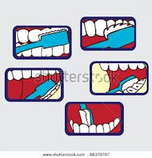
brushingOral hygiene is an important or rather essential aspect of general health. The oral cavity being an open moist area, it is prone to infection both from external and internal sources.

The innermost third of the oral cavity forms the first level of the throat. (This is a general description) the ear, nose and mouth open into it so it essentially behaves like a large pool; this makes it a convenient location for infection to breed.

Major oral problems are cavities, gum diseases, oral cancer, infections (things like Herpes,) injuries and some hereditary lesions.

The outcome of poor oral hygiene.

  • High sugar levels in the diet can lead to dental caries.
  • Smoking can lead to periodontal disease  and contribute to oral cancer
  • Systemic disorders like diabetes can aggravate caries
  • Chronic unattended caries can lead to systemic disease.
  • Hormonal changes during pregnancy, menstruation can cause a change in the oral flora, resulting in fungal infections.

We are very familiar with dealing with oral hygiene products like toothpaste, mouthwash, floss etc. we have also heard of clove, cinnamon as home remedies. Here is a sharing from Himalaya herbal healthcare

  1. Lavanga (Synzgium Aromaticum) commonly called clove.  What clove does it is produces an alkaloid that inhibits the growth of pathogens that can cause gum disorder. It is anti-inflammatory, anti-microbial and immunostimulant in nature.
  2. Pilu (Salvadora Persica) or the mustard tree.  Known as meswak it is a popular dentifrice.  This is an analgesic, antibacterial, antimycotic, astringent and also reduces plaque formation, making it effective in counteracting toothache, bad-breathe and preventing caries.
  3. Putiha (Mentha Piperita) or the peppermint.  The essential oil from Mentha Piperita is peppermint. This colourless oil (it is sometimes pale yellow in colour) has a distinct strong odour and is very effecting in fighting bad-breathe. It is antiseptic and antimicrobial, making it a must in every dentifrice, mouth wash and breathe freshener

Personally in my clinic I had good results with HiOra-K in the case of bleeding gums. This is toothpaste. Particularly in patients who are prone for gingivitis I like to use this as prophylactic option. HiOra-K mouthwash in severe halitosis.


.

Trichoptilosis

tresamme-2Right as a teenager, I had this issue of feeling things crawl on my skin; this would be followed by terrible itching and rashes. My back and forehead were particularly susceptible. After the burns it has turned worse.

I remember visiting the skin department at KMC Manipal and the doctor diagnosing Trichoptilosis

Trichoptilosis   has its origin in Greek.

Tricho= hair

Ptilosis== arrangement of feathers.

Together it describes fraying or splitting of the hair shaft due stress.

Hair splits when it becomes dry or damaged and the only way to get rid of this is to get the hair trimmed. But preventive care can be done.

One needs to find out if the hair is split only in the end or has multiple splits on the length of the same strand. Trimming the split ends is a good idea

  • The trimming should be done at least an inch above the split.
  • Catching the split early is crucial else the split could increase in length right up to the scalp.

Periodic trimming of the hair will rid it off the split end and promote growth and as well as giving a healthy look to the hair. Once the split end is trimmed out, it products should be used to prevent its split.

Products that claim to seal the split end do not really reverse the damage; they only make the hair appear healthier. But these products do prevent future split ends.

Uses of deep conditioning once a week, oil, and hair masks are all great ways to maintain hair health.

Like grandma said don’t brush wet hair, as the hair is more fragile?

Constant brushing and styling, frequent washing and using hot tools could all damages the hair making it brittle and more prone to splits.

Before drying or straightening the hair it’s a great idea to use heat protection serum, it protects the hair from getting damaged due to heat.

About a fortnight ago I received my trail pack from Tresammè, for the Tresammè experience with my knowledge of Trichoptilosis I was really sceptic. With the first wash there was kind of bounce it took two days for the hair to become brittle enough to irritate the skin.

Before the second wash I had ,had a hot oil head massage, so the hair after the wash was very limp.

post tresamme  hair style
post tresamme hair style

After the third wash I followed it with a leave on hair mask. Believe me for nearly four days I was free of the teasing from the hair tip. I did not have to cut my hair, or tie it up like I do every summer to prevent skin irritation.

The Tresammè split remedy system http://www.tresemme.com/product/category/332737/split-remedy  I realized gently cleanses and moisturizer the hair, so it becomes manageable and looks healthy. The unique reconstruction complex binds the split ends with continued use it fixes the damage as soon as it occurs. As for reducing the split ends up to 80% I could not get it graded by my hair stylist.  But of course my hair is returning to its fresh soft smooth appearance and is much easier to style.

Tresammè, of course had sent the shampoo with the conditioner but the split end sealing serum that really got me hooked.

I’m glad I tried it out.

THE TOBACCO EPIDEMIC

smokerrsSharing from tobacco advocacy workshop conducted by VHA-Goa and GoaCan/

The Global Scene

Tobacco is largest killer worldwide. 1-10 deaths worldwide are tobacco associated.  80% of these being in developing countries.

29% of the world population is addicted to smoking. One might argue that it is not really very significant. What makes it significant is the tobacco abuser debuts at a young age of 13- 15. This abuse lowers his life span and he exits but his place is taken by the next addict. That is to say 29% might be constant and legible but within the 29% there is constant new infusion. Of the tobacco abusers, 48% of men and 10.3%of women are smokers. The rest use smokeless tobacco. 84% of the tobacco abusers are in the developing countries.

In the 20th century 100 million deaths were caused by tobacco it is to hit 1 a billion in 21st century and by 2030 70% of the tobacco deaths will be developing countries.

The Indian Scene

India records 9 lakh tobacco and tobacco associated deaths. 2200 Indians die every day in a tobacco related death. And each day 5500 Indian youth start smoking—this makes the 29% relevant.

India reports the largest number of oral cancers. And 40% of the Indian cancer etiology is tobacco associated.

The Demography of Indian tobacco Users:

57% male and 11% females use tobacco in one form or the other.

Women chew tobacco more than smoke. This is more prevalent in the rural areas as compared to urban.

NE India records maximum tobacco abuse.

40.3% on Indians suffer from secondary tobacco inhalation.

Tobacco And Youth

Tobacco in the Indian context becomes very complicated as it presents itself in different form. Though smoking is the most widely acknowledged form.

37% of children start tobacco use by the age of 10.smoking forms 4.2% of this and more boys smoke than girls.11.9% of students use other tobacco products.

Various forms of tobacco presentation

Smoke presentation—cigarette, beedi, cigars, dhumti, cheroot, chutta, pipes, chillum, hooklis, hookah.

Smokeless presentation – gutka, zarda khaini, paan masala, paan masala with tobacco, tobacco water, meeta mawa, snuff, gul—applied on the teeth, gudhka—toothpaste, dentobac—toothpaste  Mishri—applied on the tooth.

Chemical Analysis of The Tobacco Concoction.

Tobacco contains psycho active component nicotine and nine other alkaloids. Nicotine is highly addictive. It causes increase in heart rate, blood pressure, and increase in blood fats. 4800 chemicals are found in a burning cigarettes, 69 of the identified ones are carcinogenic or the ones that cause cancer.

Nicotine à acts on the brain to release dopamine. Dopamine causes euphoria, and speeds metabolism. Speedy metabolism reduces hunger. The dopamine induced high drops causing a low that manifests as irritability and decreased concentration level. This in turn calls for the need for a fix. Or the next nicotine intake.

The short-term ill effects of this are—bad breathe, body odour, hair odour, stained fingers and teeth, tooth decay, breathlessness and sometimes even asthma.

Long-term adverse effect being – heart disease, reproductive disorders leading to impotency, cancer, respiratory embarrassment, stroke, neonatal deformities  that is deformities in a new-born child. Shrinking of the brain resulting in cognitive dysfunction, Alzheimer’s, cataract and loss of vision.

What is second-hand smoking?

Second hand smoking is the smoke inhaled by a non-smoker by being in the vicinity of a smoker. This is the mixture of exhaled smoke by a smoker along with the smoke emitted by the burning cigarettes. 30% increase of heart disease, 30% increase risk of lung cancer, respiratory illness asthma attacks, ear problems and if the expectant mother is a smoker then there could SIDS

And now third hand smoking

The invisible yet toxic brew of gaseous and particles clinging to the smokers hair and clothing. This lingers longer than the visible huff smoke trapped in the cushion covers curtains etc. and is a great threat to the children’s health. The residue that settles on the surface touched by the secondary smoke, (combination of exhaled smoke and burning cigarettes) contains heavy metals, carcinogens and sometimes even radioactive materials. This gets can get ingested into the system of babies, causing health issues for life.

References:

  • Effective strategies for tobacco control manual for NGO’s HRIDAY N.Delhi.
  • Tobacco control laws a resource manual HRIDAY N.Delhi
  • Voluntary Health Association of India – Website.